Name: Caroline (Lindy) Rae
Position/s:
Chair of Brain Sciences, UNSW
Senior Principal Research Scientist, NeuRA
Director (Research) NeuRA Imaging
Secretary, International Society for Neurochemistry (ISN) 2019-
How has the Neuroscience, Mental Health and Addiction Theme and CAG enabled you to develop your research interests?
Some problems in neuroscience require an interdisciplinary approach if they are to be properly addressed. The neuroscience, mental health and addiction theme, along with the requirement for investigators from multiple schools and/or faculties sets the landscape for fruitful collaborations. For example, the project of ours that has recently been funded involves researchers with expertise in neurology, biomedical engineering, signal processing, modelling, magnetic resonance imaging, EEG and physiology. The CAG also provides an opportunity to develop projects that are highly innovative and therefore seen as risky by mainstream grant agencies. This scheme allows us to develop such projects to the point where they sufficiently mature to complete for funding against more accepted approaches.
Your project, Investigation of the utility of tissue conductivity imaging for detection, location and management in epilepsies was successful in the 2020 round of Theme and CAG collaborative research seed funding. Can you please tell us about the project?
Knowledge of the location and frequency of ictal discharges is crucial to manage and treat seizures. Measurement of the degree and source of ictal discharges is currently undertaken by attaching electrodes to the surface of the skull, implanting them on the surface of the brain or inserting deep brain electrodes into the brain to record from deeper sources. We have developed and optimised a new, non-invasive MR imaging method, Magnetic Resonance Electrical Properties Tomography (MREPT) which has the potential to reliably and repetitively detect changes in brain activity and which could prove useful for detection and location of epileptic foci. This approach is completely novel, and is untried in this patient population.
Here, in a new collaboration, we will recruit selected and appropriate participants with diagnosed focal (lesion and non-lesion related) epilepsy or absence epilepsy. We will obtain conductivity images from these participants along with in-scanner EEG measurements. This will demonstrate whether or not this approach can be of use in the detection and management of seizures and provide proof of concept data for further grant applications and development.
What impact do you imagine the project will have?
This project gives the opportunity to directly measure electrical properties of brain tissue, i.e. to quantify electrical excitability, the fundamental property that is altered in epilepsy. This could possibly lead to an improvement in both sensitivity and specificity, since small conductivity changes (epileptic discharges) that do not affect neuronal oxygen consumption may be detected, while purely hemodynamic changes, which are picked up by existing imaging methods such as BOLD (Blood oxygen level dependent) imaging will be excluded. The method may allow non-invasive detection of epileptic discharges that are currently only detected via insertion of electrodes into the brain. It may also allow identification of epileptic foci to improve outcomes in cases requiring surgical focus ablation.
How will the project support new collaborations?
This project will allow us to show proof of concept for this new approach but once that is established there are a plethora of applications for this method. There is scope for much further investigation in epilepsy as well as for the application of the imaging method to a range of disorders and physiological investigations.
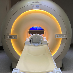
The 3T scanner at NeuRA Imaging is a funded facility of the Australian National Imaging Facility and is completely open access. This means that any researcher can access and use the facility. Conductivity imaging is just one of the capabilities of our new scanner and we encourage researchers to contact us about using it.
@NeuRAImaging

